- O repozytorium
- Kolekcje
- Indeksy
- Historia przeglądania
-
Repozytoria RCIN
-
 INSTYTUT ARCHEOLOGII I ETNOLOGII POLSKIEJ AKADEMII NAUK
-
 INSTYTUT BADAŃ LITERACKICH POLSKIEJ AKADEMII NAUK
-
 INSTYTUT BADAWCZY LEŚNICTWA
-
 INSTYTUT BIOLOGII DOŚWIADCZALNEJ IM. MARCELEGO NENCKIEGO POLSKIEJ AKADEMII NAUK
-
 INSTYTUT BIOLOGII SSAKÓW POLSKIEJ AKADEMII NAUK
-
 INSTYTUT CHEMII FIZYCZNEJ PAN
-
 INSTYTUT CHEMII ORGANICZNEJ PAN
-
 INSTYTUT FILOZOFII I SOCJOLOGII PAN
-
 INSTYTUT GEOGRAFII I PRZESTRZENNEGO ZAGOSPODAROWANIA PAN
-
 INSTYTUT HISTORII im. TADEUSZA MANTEUFFLA POLSKIEJ AKADEMII NAUK
-
 INSTYTUT JĘZYKA POLSKIEGO POLSKIEJ AKADEMII NAUK
-
 INSTYTUT MATEMATYCZNY PAN
-
 INSTYTUT MEDYCYNY DOŚWIADCZALNEJ I KLINICZNEJ IM.MIROSŁAWA MOSSAKOWSKIEGO POLSKIEJ AKADEMII NAUK
-
 INSTYTUT PODSTAWOWYCH PROBLEMÓW TECHNIKI PAN
-
 INSTYTUT SLAWISTYKI PAN
-
 SIEĆ BADAWCZA ŁUKASIEWICZ - INSTYTUT TECHNOLOGII MATERIAŁÓW ELEKTRONICZNYCH
-
 MUZEUM I INSTYTUT ZOOLOGII POLSKIEJ AKADEMII NAUK
-
 INSTYTUT BADAŃ SYSTEMOWYCH PAN
-
 INSTYTUT BOTANIKI IM. WŁADYSŁAWA SZAFERA POLSKIEJ AKADEMII NAUK
-

Obiekt
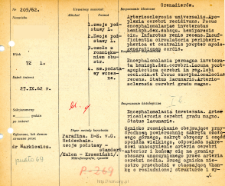
Tytuł: File of histopathological evaluation of nervous system diseases (1962) - nr 205/62
Twórca instytucjonalny:
Department of Experimental and Clinical Neuropathology MMRI
Współtwórca:
Miejsce wydania:
Opis:
Clinical, anatomical and histological diagnosis
Abstrakt:
Autopsy examination of 72-year-old patient was performed. Neuropathological evaluation in light microscopy was based on brain paraffin sections stained with Hematoxylin-eosin, van Gieson, and Heidenhain method.A foci of malacia involving the medial surface of the superior frontal gyrus, the cingulate gyrus, and part of the corpus callosum was found.The edge of the focus transitioned into surrounding tissue with a loosened structure and prominent diffuse glial proliferation. Glial reactions were not limited to glial cell densification (mainly macro and oligodendroglial), mast cells were also present. In the central part of the foci, irregular fields of macrophages loaded with brown blood stain were observed, which penetrated the surrounding tissue and separated fields of damaged cortex and white matter, sometimes with thrombotic necrosis or incomplete necrosis.Connective tissue organization was observed throughout the focus in van Gieson staining. In the putamen and caput nuclei caudati, single larger perivascular necroses with macrophages and small-cell infiltration were found among a plaque-like thickening of oligodendroglium. In addition, numerous small vascular lacunae were seen, not only in the basal ganglia but also in the white matter at the prominent atherosclerotic lesions with vitrification and fibrosis. Parenchymal damage in the form of cellular defects and ischemic changes, more extensive in the closest vicinity of the focus, leading to the obliteration of the stratified structure of the cortex and diffuse thickening of cellular glia.Histological diagnosis: Encephalomalacia inveterata. Arte riosclerosis cerebri gradu magno. Status lacunaris
Format:
Identyfikator zasobu:
Język:
Język streszczenia:
Prawa:
Creative Commons Attribution BY 4.0 license
Zasady wykorzystania:
Copyright-protected material. [CC BY 4.0] May be used within the scope specified in Creative Commons Attribution BY 4.0 license, full text available at: ; -
Digitalizacja:
Mossakowski Medical Research Institute PAS
Lokalizacja oryginału:
Library of the Mossakowski Medical Research Institute PAS
Dofinansowane ze środków:
Dostęp:
Kolekcje, do których przypisany jest obiekt:
- Instytut Medycyny Doświadczalnej i Klinicznej PAN > Dane Badawcze > Kartoteka oceny histopatologicznej chorób układu nerwowego Zakładu Neuropatologii
Data ostatniej modyfikacji:
29 mar 2022
Data dodania obiektu:
1 paź 2021
Liczba pobrań / odtworzeń:
201
Wszystkie dostępne wersje tego obiektu:
https://rcin.org.pl/imdik/publication/251077
Wyświetl opis w formacie RDF:
Wyświetl opis w formacie RDFa:
Wyświetl opis w formacie OAI-PMH:
| Nazwa wydania | Data |
|---|---|
| opis nr 205/62 | 29 mar 2022 |


