- O repozytorium
- Kolekcje
- Indeksy
- Historia przeglądania
-
Repozytoria RCIN
-
 INSTYTUT ARCHEOLOGII I ETNOLOGII POLSKIEJ AKADEMII NAUK
-
 INSTYTUT BADAŃ LITERACKICH POLSKIEJ AKADEMII NAUK
-
 INSTYTUT BADAWCZY LEŚNICTWA
-
 INSTYTUT BIOLOGII DOŚWIADCZALNEJ IM. MARCELEGO NENCKIEGO POLSKIEJ AKADEMII NAUK
-
 INSTYTUT BIOLOGII SSAKÓW POLSKIEJ AKADEMII NAUK
-
 INSTYTUT CHEMII FIZYCZNEJ PAN
-
 INSTYTUT CHEMII ORGANICZNEJ PAN
-
 INSTYTUT FILOZOFII I SOCJOLOGII PAN
-
 INSTYTUT GEOGRAFII I PRZESTRZENNEGO ZAGOSPODAROWANIA PAN
-
 INSTYTUT HISTORII im. TADEUSZA MANTEUFFLA POLSKIEJ AKADEMII NAUK
-
 INSTYTUT JĘZYKA POLSKIEGO POLSKIEJ AKADEMII NAUK
-
 INSTYTUT MATEMATYCZNY PAN
-
 INSTYTUT MEDYCYNY DOŚWIADCZALNEJ I KLINICZNEJ IM.MIROSŁAWA MOSSAKOWSKIEGO POLSKIEJ AKADEMII NAUK
-
 INSTYTUT PODSTAWOWYCH PROBLEMÓW TECHNIKI PAN
-
 INSTYTUT SLAWISTYKI PAN
-
 SIEĆ BADAWCZA ŁUKASIEWICZ - INSTYTUT TECHNOLOGII MATERIAŁÓW ELEKTRONICZNYCH
-
 MUZEUM I INSTYTUT ZOOLOGII POLSKIEJ AKADEMII NAUK
-
 INSTYTUT BADAŃ SYSTEMOWYCH PAN
-
 INSTYTUT BOTANIKI IM. WŁADYSŁAWA SZAFERA POLSKIEJ AKADEMII NAUK
-

Obiekt
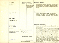
Tytuł: File of histopathological evaluation of nervous system diseases (1966) - nr 35/66
Twórca instytucjonalny:
Department of Experimental and Clinical Neuropathology MMRI
Współtwórca:
Opis:
Clinical, anatomical and histological diagnosis
Abstrakt:
Histological diagnosis: Focus metastaticus carcinomatis adcerebellum. Autopsy examination of 64-year-old patient was performed. Neuropathological evaluation in light microscopy was based on brain paraffin sections stained with H-E and Van Gieson's method.A metastatic tumor focus of adenocarcinoma type was seen in the cerebellum in the cortical-subcortical region. The tumor was well defined from its surroundings, the neoplastic tissue surrounded a field of white matter in which malacia was observed.The tumor itself was richly cellular, tubular in structure, and quite profusely vascularized. The vessels of the tumor had fibrous changes within the walls. Histological diagnosis: Focus metastaticus carcinomatis ad cerebellum.
Format:
Identyfikator zasobu:
Język:
Język streszczenia:
Prawa:
Creative Commons Attribution BY 4.0 license
Zasady wykorzystania:
Copyright-protected material. [CC BY 4.0] May be used within the scope specified in Creative Commons Attribution BY 4.0 license, full text available at: ; -
Digitalizacja:
Mossakowski Medical Research Institute PAS
Lokalizacja oryginału:
Library of the Mossakowski Medical Research Institute PAS
Dofinansowane ze środków:
Dostęp:
Kolekcje, do których przypisany jest obiekt:
- Repozytorium Cyfrowe Instytutów Naukowych > Dane i obiekty naukowe > Nauki medyczne > Kartoteka przypadków medycznych
- Repozytorium Cyfrowe Instytutów Naukowych > Kolekcje Partnerów > Instytut Medycyny Doświadczalnej i Klinicznej PAN > Dane Badawcze > Kartoteka oceny histopatologicznej chorób układu nerwowego Zakładu Neuropatologii
Data ostatniej modyfikacji:
1 lut 2022
Data dodania obiektu:
21 cze 2021
Liczba pobrań / odtworzeń:
43
Wszystkie dostępne wersje tego obiektu:
https://rcin.org.pl/publication/229193
Wyświetl opis w formacie RDF:
Wyświetl opis w formacie RDFa:
Wyświetl opis w formacie OAI-PMH:
| Nazwa wydania | Data |
|---|---|
| opis nr 35/66 | 1 lut 2022 |