Object
Title: File of histopathological evaluation of nervous system diseases (1963) - nr 16/63
Institutional creator:
Department of Experimental and Clinical Neuropathology MMRI
Contributor:
Place of publishing:
Description:
Clinical, anatomical and histological diagnosis
Abstract:
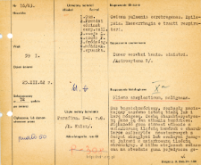
Histological diagnosis: Glioma anaplasticum, malignans.Autopsy examination of 59-year-old patient was performed. Neuropathological evaluation in light microscopy was based on brain paraffin sections stained with Hematoxylin-eosin and van Gieson.A large-cell tumor was found, infiltrating both the white matter and the cerebral cortex. The tumor was characterized by two cellular weaves. Most of the tumor was composed of a moderate amount of tumor astrocyte cells with large, round or oval, light nuclei with a moderate amount of chromatin. Single gemistocytes were visible in several places, especially around the periphery of the tumor. Clusters of neoplastic cells were found in the background of the above-described tumor. Cell nuclei also resembled neoplastic astrocytes, extremely anaplastic, most of the nuclei were highly hyperchromatic. Numerous cellular mitoses were visible. Moderate tumor vascularization in the less compact part was visible; larger with a dense network of capillaries in the areas of higher cell density. Vessels were generally unchanged, some with fibrotic and calcified features. Quite a lot of calcium deposits around the cysts and in the tissue were observed. In the medulla oblongata, some of the nerve cells were characterized by fatty degeneration, with an invisible nucleus and tigrolysis. No reticulin fibers were found in the Perdrau staining.
Format:
Resource Identifier:
Language:
Language of abstract:
Rights:
Creative Commons Attribution BY 4.0 license
Terms of use:
Copyright-protected material. [CC BY 4.0] May be used within the scope specified in Creative Commons Attribution BY 4.0 license, full text available at: ; -
Digitizing institution:
Mossakowski Medical Research Institute PAS
Original in:
Library of the Mossakowski Medical Research Institute PAS
Projects co-financed by:
Access:
Object collections:
- Digital Repository of Scientific Institutes > Scientific data and objects > Medical science > File of medical cases
- Digital Repository of Scientific Institutes > Partners' collections > Mossakowski Medical Research Institute PAS > Failes > File of histopathological evaluation of the nervous system diseases of the Department of Neuropathologu
Last modified:
Feb 21, 2022
In our library since:
Sep 9, 2021
Number of object content downloads / hits:
29
All available object's versions:
https://rcin.org.pl/publication/246226
Show description in RDF format:
Show description in RDFa format:
Show description in OAI-PMH format:
| Edition name | Date |
|---|---|
| opis nr 16/63 | Feb 21, 2022 |

 INSTYTUT ARCHEOLOGII I ETNOLOGII POLSKIEJ AKADEMII NAUK
 INSTYTUT BADAŃ LITERACKICH POLSKIEJ AKADEMII NAUK
 INSTYTUT BADAWCZY LEŚNICTWA
 INSTYTUT BIOLOGII DOŚWIADCZALNEJ IM. MARCELEGO NENCKIEGO POLSKIEJ AKADEMII NAUK
 INSTYTUT BIOLOGII SSAKÓW POLSKIEJ AKADEMII NAUK
 INSTYTUT CHEMII FIZYCZNEJ PAN
 INSTYTUT CHEMII ORGANICZNEJ PAN
 INSTYTUT FILOZOFII I SOCJOLOGII PAN
 INSTYTUT GEOGRAFII I PRZESTRZENNEGO ZAGOSPODAROWANIA PAN
 INSTYTUT HISTORII im. TADEUSZA MANTEUFFLA POLSKIEJ AKADEMII NAUK
 INSTYTUT JĘZYKA POLSKIEGO POLSKIEJ AKADEMII NAUK
 INSTYTUT MATEMATYCZNY PAN
 INSTYTUT MEDYCYNY DOŚWIADCZALNEJ I KLINICZNEJ IM.MIROSŁAWA MOSSAKOWSKIEGO POLSKIEJ AKADEMII NAUK
 INSTYTUT PODSTAWOWYCH PROBLEMÓW TECHNIKI PAN
 INSTYTUT SLAWISTYKI PAN
 SIEĆ BADAWCZA ŁUKASIEWICZ - INSTYTUT TECHNOLOGII MATERIAŁÓW ELEKTRONICZNYCH
 MUZEUM I INSTYTUT ZOOLOGII POLSKIEJ AKADEMII NAUK
 INSTYTUT BADAŃ SYSTEMOWYCH PAN
 INSTYTUT BOTANIKI IM. WŁADYSŁAWA SZAFERA POLSKIEJ AKADEMII NAUK


